Osaka University Researchers Use CNN to Predict Quality of Hypothalamus-Pituitary Organoids from Early Surface Morphology
In a groundbreaking advancement for regenerative medicine, researchers at Osaka University have developed a convolutional neural network (CNN) that predicts the quality of hypothalamus-pituitary organoids derived from human induced pluripotent stem cells (iPSCs) with remarkable accuracy. Led by Professor Takuya Yamamoto and Assistant Professor Ryusaku Matsumoto from the Department of Life Science Frontiers, the team’s innovative model leverages early-stage surface morphology to forecast long-term differentiation outcomes, offering a non-invasive solution to a critical challenge in organoid research. Published in Communications Biology on December 6, 2024, this work promises to enhance the efficiency and reproducibility of organoid-based studies, with significant implications for clinical applications.
The Challenge of Organoid Variability
Hypothalamus-pituitary organoids, three-dimensional (3D) cell cultures mimicking the pituitary gland and hypothalamus, are pivotal for studying neurodevelopmental processes and advancing regenerative therapies, such as transplantation for hormone-related disorders. However, their development is hindered by protracted culture periods—often exceeding two months—and significant variability in quality. This variability stems from the heterogeneous cell populations inherent in 3D cultures, making it difficult to maintain consistent differentiation efficiency. Traditionally, researchers rely on empirical assessments or molecular markers that manifest late in the process, leading to resource-intensive protocols with uncertain outcomes.
A Novel CNN-Based Approach
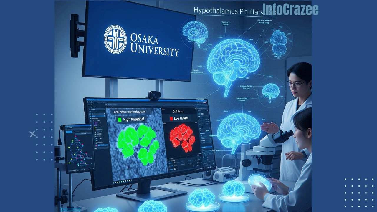
The Osaka University team addressed this bottleneck by developing a CNN model trained on phase-contrast images captured during the early stages of organoid development. Achieving an impressive 79% accuracy, the model predicts pituitary cell differentiation at day 40 using images from as early as day 9. Unlike previous approaches that required concurrent training and evaluation, this model forecasts long-term outcomes based solely on early morphological features, marking a significant leap in predictive capability.
Key Features of the Model
- Early Prediction: By analyzing phase-contrast images at day 9, the CNN identifies surface morphology—specifically budding patterns and texture—as critical predictors of organoid success. Successful organoids exhibit small budding areas and slightly rough surfaces, while failed organoids often display smooth or irregularly rough textures associated with mislocalized neural or retinal cells.
- Grad-CAM Visualization: The team employed Gradient-weighted Class Activation Mapping (Grad-CAM) to understand the model’s decision-making process. This technique highlighted surface morphology as a key determinant, revealing that structural cues precede molecular differentiation markers, enabling early intervention in the culture process.
- Superior Performance: The CNN outperformed human experts, who achieved lower accuracy (approximately 67%) in morphological assessments, demonstrating the model’s ability to extract subtle features invisible to the trained eye.
Technical Insights
The model’s robustness was enhanced by high-resolution imaging and large training datasets, though deviations in focal position during imaging reduced reliability, underscoring the need for standardized protocols. The CNN also demonstrated versatility by predicting outcomes in cell lines without fluorescent reporters (e.g., RAX::VENUS), suggesting its potential for broad clinical applications, such as organoid screening for transplantation.
Implications for Regenerative Medicine
This breakthrough has far-reaching implications for organoid research and regenerative medicine:
- Improved Efficiency: Early prediction allows researchers to terminate failing cultures, saving time and resources. This is critical in high-throughput settings where scalability is a priority.
- Enhanced Reproducibility: By identifying viable organoids early, the model reduces variability, improving the reliability of experimental outcomes.
- Clinical Translation: The non-invasive nature of the CNN, relying on phase-contrast imaging without complex fluorescent reporters, makes it adaptable for clinical environments, such as quality control in organoid-based therapies for pituitary disorders.
- Broader Applications: The methodology could extend to other organoid types, such as retinal or cerebral organoids, where early morphological assessment is equally critical.
Real-World Impact
The Osaka University model addresses a key bottleneck in organoid research, where long culture periods and inconsistent outcomes have limited scalability. For example, in regenerative medicine, high-quality hypothalamus-pituitary organoids are essential for developing therapies for conditions like hypopituitarism. By enabling early, non-invasive assessment, the CNN could streamline the production of transplantable organoids, potentially reducing costs and accelerating clinical trials. Additionally, the model’s ability to outperform human experts highlights the power of AI in augmenting scientific discovery, particularly in complex biological systems.
Challenges and Future Directions
Despite its promise, the approach faces challenges:
- Imaging Standardization: Variations in imaging protocols, such as focal position, can impact model reliability, necessitating standardized equipment and procedures.
- Data Requirements: Large, high-quality datasets are critical for training robust CNNs, which may pose challenges for smaller labs.
- Generalization: While the model is robust across certain cell lines, further validation is needed to ensure applicability to diverse iPSC sources.
Looking ahead, the team plans to integrate additional data types, such as time-series imaging or molecular profiles, to enhance prediction accuracy. Combining CNNs with other AI architectures, like Vision Transformers, could further improve performance, as suggested by related studies.
Conclusion
The work by Yamamoto and Matsumoto at Osaka University represents a significant advancement in organoid research, leveraging AI to overcome longstanding challenges in hypothalamus-pituitary organoid development. By predicting quality from early surface morphology with 79% accuracy, their CNN model offers a non-invasive, efficient, and scalable solution for regenerative medicine. As healthcare continues to embrace AI-driven innovation, this breakthrough paves the way for more reliable organoid production, with the potential to transform research and clinical applications. For researchers and clinicians, adopting such AI tools will be critical to unlocking the full potential of organoids in addressing complex medical challenges.